ADVANCED VISUALIZATION

Terarecon Intuition

KLAS Category Leader & #1 US Market Shareholder for 3D Imaging

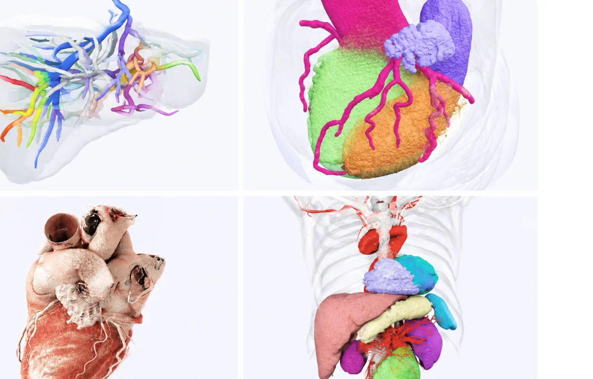
TeraRecon consistently leads the advanced visualization category of the industry's leading independent technology analyst. Offering advanced 3D and 4D medical imaging post-processing, TeraRecon is widely considered the gold standard in cardiac and vascular imaging.

TeraRecon is continually innovating to bring the industry the latest in AV capabilities! The latest version of our best-in-class advanced visualization software is packed with AI productivity and novel cardiac MR medical imaging workflows. With over 200 enhancements, Intuition 4.10 delivers unmatched precision for cardiac CT and MR, and is now available on a robust cloud-based SaaS platform.

Terarecon Intuition

Intuition Packages

2D Analysis

2D Analysis

  • 2D workflow, tools, and measurements
  • Available within the 2D workflow or when evaluating 2D images anywhere in Intuition

Experience the practicality and precision of TeraRecon's 2D Analysis solution, designed to integrate seamlessly within the Intuition platform. The 2D Analysis solution offers an array of tools and measurements, specifically tailored for evaluating 2D images within the dedicated 2D workflow or while analyzing 2D images anywhere in Intuition.

This efficient and reliable solution simplifies the diagnostic process, enhancing workflow and providing accurate results for greater diagnostic confidence. With its intuitive design, the 2D Analysis solution ensures streamlined integration with the Intuition platform, making it an essential tool for medical professionals seeking improved performance and productivity.
Autobatch

Autobatch

  • 2D batch output - reformation of image data into alternative planes
  • Create a derived series with any number of images, FOV, slab thickness, slice spacing, and rendering mode including MIP

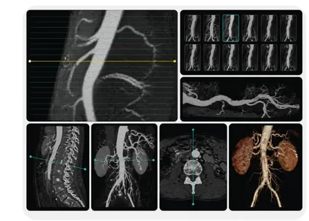
Autobatch utilizes Aquarius APS to provide users with an advanced pre-processing engine that is designed to give a 2D batch output - enabling the reformation of image data into alternative planes or the creation of movies. The system can be configured to create a derived series with any number of images, along with adjustable field of view, slab thickness, slice spacing, and rendering modes, including MIP. Processed images will be appended as additional series to the original studies.

Body Fusion

Body Fusion

  • Registration fusion
  • Subtraction
  • CT, MR, PET, SPECT
  • Motion-correction
  • Min, max, mean, standard deviation, standard uptake values (SUV)
  • Findings viewer and follow-up
  • Mirada XD CT-MR image registration for PET/CT to MR fusion

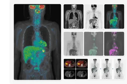
Body Fusion supports volume registration and fusion, aligning and comparing 2D and 3D images from CT, MR, PET, or SPECT data sets for anatomical reference and quantitative study. It offers automatic registration and motion correction, and generates subtractions to a third series. Validated findings can be stored for comparing multiple time points. Users can describe regions of interest (planar or volumetric) to obtain statistical quantities like min, max, mean, standard deviation, and SUV counts readout for PET data.

Manual registration tools are also available for registering different image sets together, and findings workflow stores captured values for side-by-side comparison. The new Mirada XD Add-On provides capabilities of an automatic image fusion solution that can be performed for rigid and deformable options. It can also provide image fusion between time points, and can be used for fusing multiple studies from different modalities, including PET/CT, SPECT/CT, MR, and CT.

CT Body

CT Body

  • Registration fusion
  • Subtraction
  • CT, MR, PET, SPECT
  • Motion-correction
  • Min, max, mean, standard deviation, standard uptake values (SUV)
  • Findings viewer and follow-up

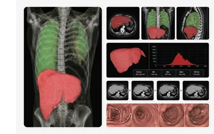
CT Body allows radiologists to perform a comprehensive review and analysis of organs in the abdomen and pelvis region. Additionally, this package enables radiologists to calculate the volume of organs or regions of interest with histogram output, and utilize exportable measurement values for follow-up comparison.

The CT Body Package also provides the ability to analyze dynamic data to support the assessment of time-dependent behavior of the image intensity or density of anatomy. Validated findings can be stored for the comparison of multiple time points. Full function colon flythrough includes automatic multi-volume side-by-side loading and viewing, fly-path creation and editing, and AquariusAPS sphere finder for sphere-like structure identification.

Dynamic image filtering with configurable filtering strengths can be engaged to improve image quality from low-dose scanning techniques. Data from dual-source scans (i.e. Dual Energy) are also supported to provide dynamic blending, improving signal-to-noise ratios and subtracting high-density structures such as bone and metal.

CT Cardiac

CT Cardiac

  • Cardiac structure segmentation and functional analysis
  • Automated centerline creation and vessel segmentation
  • Calcium scoring with multiple database options
  • Atrium and pulmonary vein analysis for EP planning
  • Pre-operative evaluation of coronary arteries for plaque and stenosis
  • Embedded geometry for pre-operative virtual stent evaluation

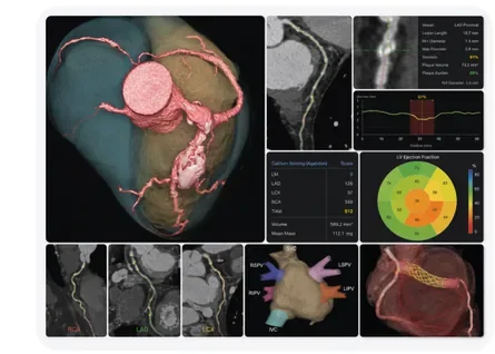
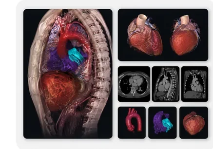
CT Cardiac provides a simplified approach to complex cardiac analysis and quantification, enabling a radiologist or cardiologist to analyze coronary vessels with zero-click centerline creation and extraction. Vascular evaluation tools include plaque and soft plaque, stenosis, curvature, tortuosity, and more. The Calcium Scoring measurement of Agatson score, volume, mean mass (mg) is also supported. In addition, the polar map from LV and RV ejection fraction can be merged with coronary marks for territory mapping of affected AHA 17 segments for heart function evaluation.

CT Chest

CT Chest

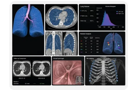
  • Automated lung segmentation
  • Lung volume and histogram analysis
  • Sphere-like structure identification
  • Comparative tracking-over-time options
  • Virtual flythrough
  • Rib Labeling

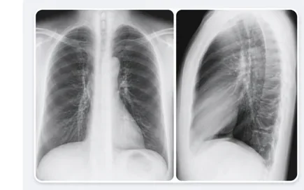
CT Chest provides pre-generated lung segmentation for lung volume measurements with volume histogram output. When image noise is a concern, dynamic image filtering with configurable filtering strengths can be engaged to improve reading efficiency. The AquariusAPS sphericity index is designed to help physicians identify and manage sphere-like structures. Validated findings can be stored for the comparison of multiple time points. Virtual flythrough guides you to visualize the airway tree to evaluate patients with a variety of airway pathologies.

CT Head and Neck

CT Head and Neck

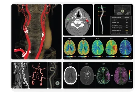
  • Automatic centerline identification simplifies stenosis measurements
  • Dual source data support
  • Time density evaluation: maps include CBF, CBV, MTT, TTP, Tmax, hypoperfusion, mismatch, and more
  • Multi-modality image fusion and image subtraction

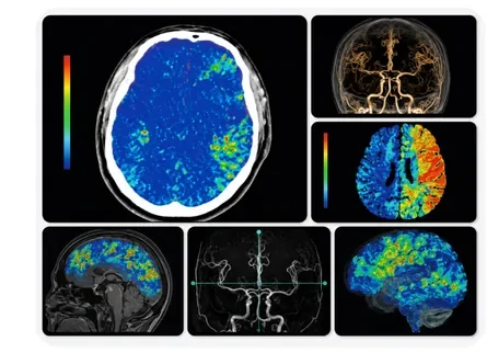
CT Head and Neck Package tools facilitate bone and vessel removal with advanced editing to support vasculature analysis including stenosis ratio, area, diameter, Min, Max, Mean, or perimeter cross-section display. In addition, radiologists can calculate the volume of organs or regions of interest with histogram output and utilize exportable measurement values for follow-up comparison.

It also provides the ability to analyze dynamic data to support the assessment of time-dependent behavior of the image intensity or density of the brain, including CBF, CBV, MTT, TTP, Tmax, hypoperfusion, mismatch, and more. Data from dual-source scans are also supported to provide dynamic blending, improve signal-to-noise ratios, and subtract high-density structures such as bone and metal. Image fusion for cross-planar image synchronization with customizable, vendor-specific map type color scales is also supported.

Data Adaptor

Data Adaptor

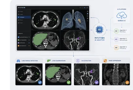
  • Allows ingestion of AI inputs from multiple algorithms
  • Delivers advanced starting point for clinical users
  • Seamlessly integrates with Intuition workflow
  • Compatible with premium first-, second-, or third-party AI algorithms

Intuition AI Adaptor enables the clinical users to experience the best of premium first-, second-, or third-party AI algorithms directly within the Intuition workflow. Included in the Intuition Titanium suite, AI Adaptor drives communication between your existing Intuition system and the Eureka AI platform to ingest available AI inputs from one or multiple algorithms to deliver an advanced starting point with near zero-click user experience.

Data Extractor

Data Extractor

  • Unlocks segmentation data potential
  • DicomSeg and RtStruct extraction efficiency
  • Data transformation for enhanced compatibility
  • Expanded data usability across platforms
  • Streamlines system integration
  • Facilitates interoperability

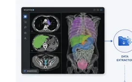
Data Extractor is a useful function designed to unlock the potential of your segmentation data. It efficiently extracts DicomSeg and RtStruct data from Intuition, transforming it into a format that can be readily utilized by other systems. This process not only expands the usability of your data, but also ensures seamless integration with various platforms. With Data Extractor, interoperability is at your fingertips, making the transition of valuable segmentation information effortless and efficient.

EVAR Workflow

EVAR Workflow

  • Pre-generated centerlines
  • User-definable planning template
  • Diameter vs. distance and cross-sectional views
  • Straightened view, diameter, and length measurements
  • Embedded vendor-specific report templates

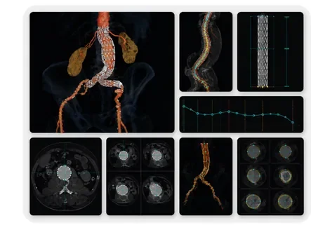
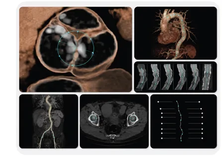
EVAR provides features including an advanced measurement protocol option, a user-definable planning template with report output and embedded instructions. Other features include AquariusAPS pre-processed centerline extraction, straightened view, diameter, length measurements, and CPR and axial renderings.

In addition, it allows for display of the diameter, distance, perimeter, cross-sectional views of vessels, and exportation of tabulated measurements. By following guided instructions, the user can complete stent-graft planning and generate reports for vendor-specific templates.

Glow Rendering

Glow Rendering

  • Photo-realistic 3D visualization
  • High-quality structure rendering

Elevate your 3D visualization capabilities with TeraRecon's Glow Rendering solution, designed to deliver photo-realistic renderings of intricate 3D structures. Our advanced rendering technology transforms complex medical data into stunning, high-quality visualizations, allowing medical professionals to better understand and interpret anatomical details.

iGENTLE

iGENTLE

  • Noise reduction management
  • Improve the effectiveness of 3D image quality
  • Improve contouring, segmentation features, and centerline accuracy

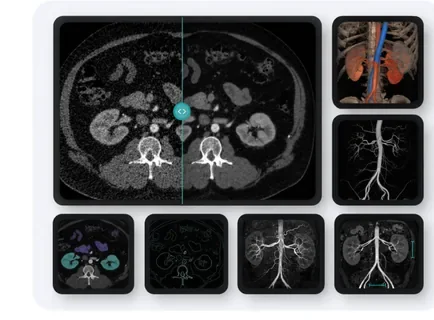
iGentle provides advanced enhancement and noise reduction management with low-dose CT exam images rendered using the APS server. TeraRecon created the iGENTLE package to reduce the effects of image noise on 3D rendering and manipulation tools supported by the software.

Such images are used internally by the software to improve the performance of the tools used by radiologists. They are not offered as any kind of substitute for the original image, however an optional iGENTLE-filtered series can be created for viewing in addition to the original data.

iGENTLE applies image-filtering algorithms that help reduce image noise and improve the 3D image quality, centerline accuracy, contouring, and segmentation features used within the system. As a result, these tools assist users in achieving their image processing objectives, even when the quality of the source image is sub-optimal.

Interventional Radiology

Interventional Radiology

  • Centerline analysis tools
  • Stent-graft planning
  • Curved planar reformation
  • Analysis and follow-up tools
  • Perspective flythrough

Interventional Radiology provides an extensive range of patented Intuition clinical and workflow tools for interventional radiologists to perform comprehensive patient analysis - including vessel stenosis calculations, aneurysm evaluation, and planning capabilities.

Zero-click pre-processed vessel centerlines maximize user productivity. Key features include Curved Planar Reformation (CPR), Straight MPR views (sMPR), Medial Axis Reformation (MAR), and perspective flythrough for endoluminal evaluation.

Left Atrial Appendage (LAA) Workflow

Left Atrial Appendage (LAA) Workflow

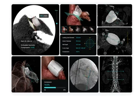
  • Guided LAA workflow
  • Optimized cardiac orientations for the LAA
  • Key measurements include landing zone, wall depth, ostium, c-arm angle, and compression measurements
  • Embedded device simulation with compression percentage
  • Fluoroscopy view with measurements and device
  • LAA report with measurements and captured images

The Left Atrial Appendage (LAA) Workflow Solution by TeraRecon revolutionizes cardiac treatment by seamlessly streamlining the entire LAA procedure. Our user-friendly, guided LAA workflow ensures a smooth and efficient process for optimal patient outcomes.

Utilizing cutting-edge technology, the system generates optimized cardiac orientations for the LAA, guaranteeing accurate visualization and assessment. Comprehensive key measurements, including landing zone, wall depth, ostium, c-arm angle, and compression measurements, are provided to enhance procedural success.

Embedded device simulation with compression percentage offers invaluable insights for informed decision-making. The fluoroscopy view with measurements and device enables real-time adjustments and monitoring, while the LAA report with measurements and captured images serves as a valuable resource for post-procedure analysis and documentation.

Liver Segmentation

Liver Segmentation

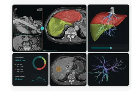
  • Semi-automated liver segmentation
  • Vascular classification options
  • Multi-cut option for pre-surgical planning
  • Dynamic image filtering with configurable filtering strengths

Liver Segmentation provides an extensive range of patented Intuition clinical and workflow tools for specialists using CT scanning for organ analysis and quantification. This package offers semi-automated liver segmentation, lesion definition with volume measurement, classification of the vasculature, and vascular centerline distance measurements for invasive treatments. The user-friendly workflow guides the user through fine-tuning segmentation both semi-automatically and manually.

Display of lesion-to-vascular relationship and lobular segmentation with a multi-cut option for pre-surgical planning is also supported.

Lung Density Analysis II

Lung Density Analysis II

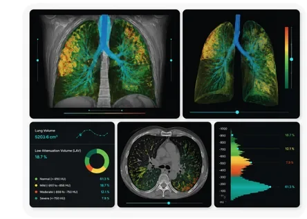
  • Lung and trachea segmentation
  • Achieve faster and better-informed care
  • Strengthen clinical decision making
  • Increase confidence

Intuition's Lung Density Analysis II workflow delivers enhanced segmentation data science and automated measurement capabilities within a guided workflow for the disease progression assessment and quantification of lung-related illness.

Lung Segmentation

Lung Segmentation

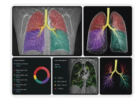
  • Lung and trachea segmentation
  • Lung and lobe volumetric analysis
  • Lung, airway, and vessel anatomical fusion
  • Treatment planning simulation
  • Low attenuation

Lung Segmentation provides an extensive range of clinical and workflow tools for thoracic and pulmonary specialists to quantify lung volumes. This package includes lobal segmentation with volume calculations, AquariusAPS sphericity index to automate lung and trachea segmentation, and low attenuation segmentation with user-configurable range values.

TeraRecon's Intuition Solution Clinical Packages include Intuition and AquariusAPS. In addition, the included findings feature can provide a side-by-side comparison of multiple time points, doubling time display.

Maxillo-Facial

Maxillo-Facial

  • Panoramic projection
  • Cross-sectional multi-planar reconstruction
  • Definable mandibular groove path

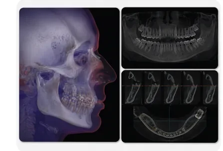
Maxillo-Facial provides an extensive range of patented clinical and workflow tools to display and manipulate dental images to support the analysis and visualization of volumetric CT datasets of the Maxillo-facial region.

The Maxillo-Facial Package applies the curved planar reformation (CPR) result to generate "panoramic" projections in various planes and thicknesses. Cross-sectional multi-planar reconstruction (MPR) may also be generated at set increments along the defined curve plane and used to obtain key measurements to aid dental implant and surgical planning.

In addition, the mandibular groove path can be displayed as an overlay to improve visualization of critical anatomy.

MR Body

MR Body

  • 2D, 3D, 4D MR image sequencing
  • MIP and MRA evaluation with centerline tools
  • Analysis and follow-up tools
  • Time-intensity ROI analysis
  • Parametric mapping of body parts such as breast or prostate
  • Kinetics, time-to-peak, time to the enhancement, and maximum slope evaluation

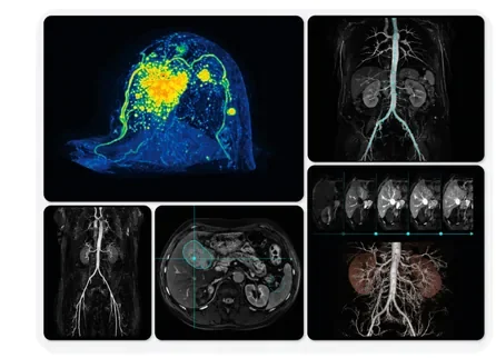
MR Body provides an extensive range of clinical and workflow tools for radiologists, internists, and organ specialists who utilize 2D, 3D, and 4D MR image sequences.

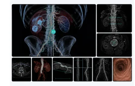
This package allows for a comprehensive yet-streamlined patient analysis of anatomy and organ function including evaluating MRA vessel analysis with a familiar centerline and editing tool. It also provides the ability to analyze dynamic data to support the time-dependent behavior of the image intensity or density of anatomy.

Radiologists can measure organ volume or regions of interest with intensity value output and utilize exportable measurement values for follow-up comparison. To support uptake curves, multiphase analysis, and tROI (time-intensity region of interest) measurement, graphical and parametric mapping displays are also provided.

Neuro Perfusion Maps

Neuro Perfusion Maps

  • Updated workflow integration
  • Enhanced Neuro maps
  • TMax, Hypoperfusion, and Mismatch results

Discover the latest advancements in neuro-imaging with TeraRecon's Neuro Perfusion Maps. This innovative solution offers an updated workflow and enhanced neuro maps for improved diagnostic accuracy and efficiency.

Featuring advanced parameters such as TMax, Hypoperfusion, and Mismatch results, our Neuro Perfusion Maps deliver valuable insights into cerebral blood flow dynamics.

MR Cardiac

MR Cardiac

  • Volumetric analysis of ejection fraction
  • LV/RV inner and outer contour detection
  • T1 Mapping, T2/T2* Mapping
  • AHA17-segment-model
  • MR flow analysis
  • MR cardiac perfusion

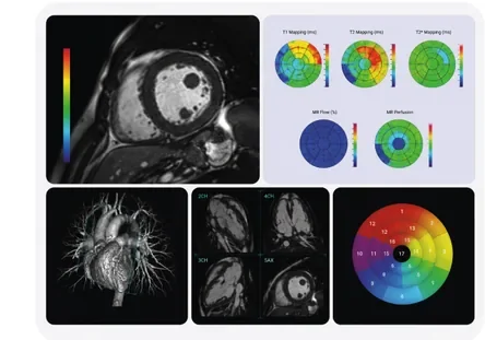
The Intuition Cardiac MR package evaluates the anatomy and physiology of the heart chamber and valves, the size and flow of blood through vessels, and the surrounding structures.

It is used to determine whether a patient suffers from cardiovascular diseases such as limited cardiac or valvular-related functional outputs.

Rib Labeling

Rib Labeling

  • Rib Labeling
  • Centerline generation
  • Fishbone image display
  • Efficient fracture detection and evaluation

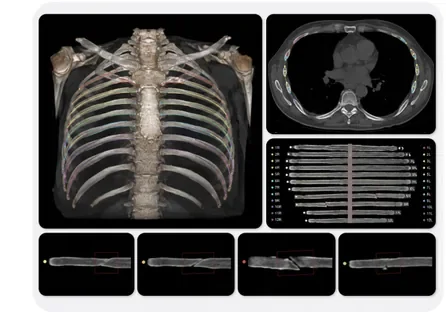
Streamline rib fracture detection and evaluation with TeraRecon's Rib Labeling solution. Our advanced technology generates centerlines for each rib and presents them in an intuitive fishbone image display, allowing medical professionals to quickly and easily identify fractures and assess rib integrity.

TAVR Workflow

TAVR Workflow

  • Aortic root segmentation and orientation
  • Centerline pre-processing and extractions
  • User-definable planning template
  • Report output
  • Automatic Measurement Protocols

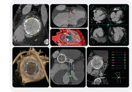
The Intuition guided workflow for TAVR aids clinicians in evaluating the aortic annulus and peripheral vascular system.

Utilizing AquariusAPS pre-processing power, the aorta and vessels are labeled and segmented for optimal efficiency. Measurement protocols guide the customer through required measurement steps through the multi-series process providing all the right measurement tools (vessel diameters, perimeter, area, distance, c-arm angles, curvature, tortuosity, and more) at the right time.

Key results and images are outputted to the defined DICOM destination.

TMVR (Mitral Valve) Workflow

TMVR (Mitral Valve) Workflow

  • Guided TMVR workflow
  • Optimized cardiac orientations for the mitral valve
  • Two-, three-, and four-chamber views, as well as oblique mitral valve view
  • Key measurements include trigon-to-trigon (TT), septal-to-lateral (SL), and intercommissural (IC) distance measurements
  • Saddle-shape or D-shape options
  • Aortomitral angle measurement
  • Embedded geometry with percent or mm offset
  • Automatic Neo-LVOT centerline and measurements
  • Summary page displays key results and images

TMVR provides tools for evaluation of the mitral valve and to provide measurements to aid the trained clinical user for TMVR treatment planning.

The workflow includes optimized layouts and image reconstructions that are helpful in annulus evaluation. This includes optimal projections (two-, three-, and four-chamber views), templates, as well as concise and user-directed steps and measurements.

These measurements and steps all culminate in the treatment planning for repair or replacement of the native valve utilizing embedded geometry that matches approved and cleared devices.

Volumetric Navigation

Volumetric Navigation

  • For 2D, 3D, and 4D viewing
  • Anatomy segmentation and volume analysis
  • Configurable workflow creation
  • Comprehensive and robust measurement tools
  • Image batching and report generation
  • Comparison views for follow-up patient review

Volumetric Navigation provides all of the most fundamental image processing tools in a single workflow.

2D, 3D, and 4D image display with MIP, MinIP, MPR, Thick MPR, and 3D rendering with flexible and comprehensive arrangements of tools, processors, templates, batch options, and more.

All results and key images may be sent to reports and exported to the preferred DICOM destination.

CONTACTS

Contact Us

For any Medical Imaging Solution requirements - RIS, PACS, Teleradiology, VNA, Ultrasound, Structured Reporting & Endoscopy Image Management, Recording & Live Streaming, Cloud Solutions, C-Arm Memory, Patient Portal, 3D Solutions, Advanced Visualization, and many more.

Get in Touch

Choose your country